

A 3D printer that can use a patient’s own cells to print skin grafts, including hair follicles and sweat glands, has claimed the James Dyson Award for student design. The group of four students from the University of Toronto will compete for a prize of $60,000 at a global final.
While still in pilot mode, the PrintAlive Bioprinter is in the process of being commercialized by MaRS Innovations in collaboration with the Innovations and Partnerships Office (IPO) of the University of Toronto, whose labs have filed two patents on the device.
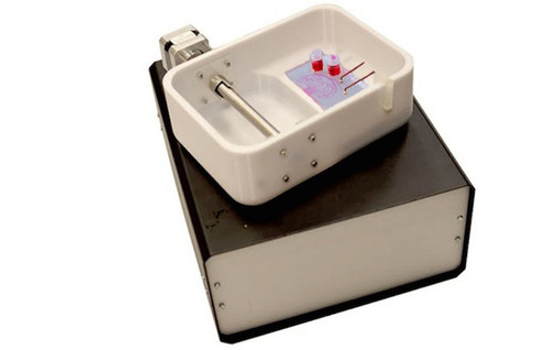
The PrintAlive Bioprinter has been able to create uniform, large-scale engineering of tissue, which could be used to treat burn patients and other injuries.
The engineers who designed the printer include associate professor Axel Guenther, associate professor Milica Radisic, and graduate students Lian Leng, Boyang Zhang, and Arianna McAllister.
“There’s a lot of interest in soft materials, particularly biomaterials,” Guenther said in a university news release. “But until now no one has demonstrated a simple and scalable one-step process to go from microns to centimeters.”
Along with producing skin cells, the machine can also produce hair follicles, sweat glands and other human skin complexities, “providing an on-demand skin graft for burn victims,” the university stated.
Because the machine uses the patient’s own cells, which are grown in a petri dish and then printed to form a bandage, it “would completely eliminate immunologic rejection, and the need for painful auto-grafting and tissue donation,” McAllister said in a statement.
3D bioprinting an up and coming market
The University of Toronto is not alone in using 3D printing technology to create human tissue. Other university researchers and private companies are developing printing technology that could someday be used to replace organs and other body parts.
San Diego-based bio-printing company Organovo expects to unveil the world’s first printed organ – a human liver – this year.
Like other forms of 3D printing, bio-printing lays down layer after layer of material – in this case, live cells – to form a solid physical entity (tissue). A major stumbling block in creating tissue continues to be manufacturing the vascular system needed to provide it with life-sustaining oxygen and nutrients.
3D printing also allows enough tissue and supportive vasculature to be constructed quickly so that enough living cells can survive to create functional tissue.
Organovo said it has also overcome the vascular issue to a degree. “We have achieved thicknesses of greater than 500 microns, and have maintained liver tissue in a fully functional state with native phenotypic behavior for at least 40 days,” said Michael Renard, Organovo’s executive vice president of commercial operations.
How it works
The University of Toronto’s PrintAlive Bioprinter 3D skin printer works by placing the patient’s cells along with other biomaterials into a micro-device, which then pushes them out through several channels. The biomaterials are then mixed, causing a chemical reaction that forms a ‘mosaic hydrogel’, a sheet-like substance compatible with the growth of cells into living tissues. The hydrogel allows the various dermis cells to be seeded in precise and controlled patterns.
The printing technique is different from others, the scientists said, because it doesn’t require an artificial, but biodegradable, scaffolding to hold the cells together. Typically, 3D bio-printing requires cells to be placed onto an artificial structure capable of supporting three-dimensional tissue formation.
The placement of the cells by the printer is so precise that the researchers were able to spell words, such as Toronto, demonstrating how they could mimic the natural placement of cells in living tissues.
The mosaic hydrogel sheets are collected around a drum, which allows the formation of layers as it turns, creating, in essence, three-dimensional, functional tissues.
“In this case, when we put the cells in the right places, we create cellular organisation quite naturally,” Leng said.
So what’s the next step?
Along with treating burn victims, the 3D-printed tissue could be used for testing therapeutic drugs, potentially eliminating the need for arduous and highly regulated animal and human trials.
The university researchers are working with a burn unit at Toronto’s Sunnybrook Hospital.
Computerworld


Subscribers 0
Fans 0
Followers 0
Followers